Aspergillus whole mount (w.m.) is the second slide in the Amscope 50PC prepared slides set. Aspergillus is a genus consisting of several hundred mold species found in various climates worldwide [1]. A mold is a fungus that grows in the form of multicellular filaments called hyphae. In contrast, fungi that can adopt a single celled growth habit are called yeasts [2].
The third slide in the Amscope 50PC prepared slides is the longitudinal section of cabbage. The individual cells of the cabbage are clearly visible in the slide.
Micrographs
Amscope 50PC Prepared Slides
This post lists all the micrographs I have done from the Amscope 50PC prepared slides.
I bought Amscope 50 piece biology prepared glass slides box along with Amscope B120B microscope to learn how to use a microscope properly and refine my techniques without worrying about making proper slides. This post list all the slides provided in the box. Each entry links to a post where I will discuss the specimen and show micrographs I recorded.
The first slide in the Amscope 50PC prepared slides is whole mount (w.m.) of ascarid eggs. Ascarid (Ascaris) is a parasitic worm also known as small intestinal roundworm. There are two species of Ascaris: Ascaris Lumbricoides and Ascaris Suum. Ascaris Lumbricoides affects humans and causes a disease known as Ascariasis and Ascaris Suum typically infect pigs [1].
This is the first prepared slide I did and you can see that they are not very clearly focused and also the white balance is not correct.
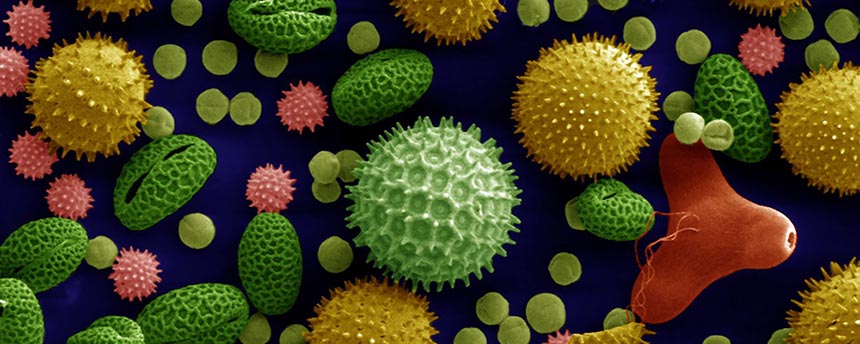
I have been interested in microscopy for very long time and the recent flurry of articles (io9.com, twitter, and telegraph.co.uk) showing very nice images further fueled my interest. These images were taken using a Scanning Electron Microscope (SEM) which can magnify objects up to 500,000 times whereas a simple light microscope can only magnify up to 1,000X or 2,000X but still the prospect of being able to look ordinary household things not visible to naked eye is really exciting to me.
Microscopy is the field of observing used to study objects, both living and dead, which are too small to be seen by naked human eye.
I live in Singapore and it is rather difficult to buy a decent and cheap microscope to start with microscopy here. I have found only one shop in Singapore selling microscopes: Astro Scientific Center Pte. Ltd (located in the Singapore Science Center). Astro Scientific Center have two good biological microscopes: Advanced Biological Microscope 500 (500$) and Professional Biological Microscope 1500 (1600$). Digital imager have to be bought separately at 266$. There are few more online stores (Carolina, Monotaro, and VWR) in Singapore which sell microscopes and other supplies but they cater to schools and other scientific institutions and are pricier for amateur microscopy.
Disappointed by the lack of local stores, initially I tried to order it online from overseas. But due to very high shipping costs, almost same as the microscope itself (microscopes are heavy!) I refrained from ordering. Then as luck would have it, my wife had to go to US for a two weeks business trip. I took advantage of the opportunity and ordered Amscope Binocular Compound Microscope B120B and 3MP USB2.0 Microscope Digital Camera MU300 from Amazon and get it shipped to her hotel :-). In addition to the microscope I order few other things as well since they are not easily available in Singapore:
All these items costs only 600$ (shipping was free!). To keep the microscope safe, I also decided to buy a Digi-Cabi DHC-100 dry cabinet from Harvey Norman for 378$. Below are some picture of the things I ordered from the Amazon.
Invalid Displayed Gallery
The microscope is of good quality and build for its price range. The optics are of good quality as well and looking directly into scope specimen appear clear and sharp. However, the 3MP camera I ordered with it is not as good as microscope. It has a slow response and thus not suitable to observe fast moving microorganism (they appear as fast moving lines on the screen). The camera also have a problem with white balance. Even on clean slides there is a slight greenish-blue tint, although the software that comes with it is great and can be used to fix the white white balance. Overall, it is still a decent camera and can take good pictures with some effort.
To test the microscope, the first slide I did was of an ant trapped between a glass slide and cover slip :-).
Ant magnified 40 times
credit for pollen image on the top: William Crochot (http://www.science-et-vie.net)
SPhdThesis is a latex document class for writing PhD thesis. I developed it while writing my PhD thesis in School of Computing (SoC), National University of Singapore (NUS). By default, it adheres to the NUS Guidelines on Format of Research Thesis Submitted For Examination (Link updated on 31/05/2014). However, it is quite easy to change it for a different guideline.
Downloading
You can download the latest version here or fork it at Github.
Getting started
The easiest way to start using SPhdThesis is to copy the example folder and start by inspecting and modifying thesis.tex. figure.tex, table.tex, algorithm.tex files show how to format figures, tables, and algorithms, respectively. thesis.pdf is an example of how a thesis looks like using SPhdThesis document class.
For more details read SPhdThesis.pdf. It describes in detail how to use the document class and provide some hints on how to customize it. It also lists some useful tools for working with latex.
Getting Help
The best place is to raise an issue on Github. Alternatively, you can use the comment form at the bottom of this post.
 magnified 40 times")
 magnified 100 times")
 magnified 100 times")
 magnified 40 times")
 magnified 100 times")
 magnified 400 times")
 magnified 1000 times")
 magnified 40 times")
 magnified 100 times")
 magnified 1000 times")
 magnified 400 times")