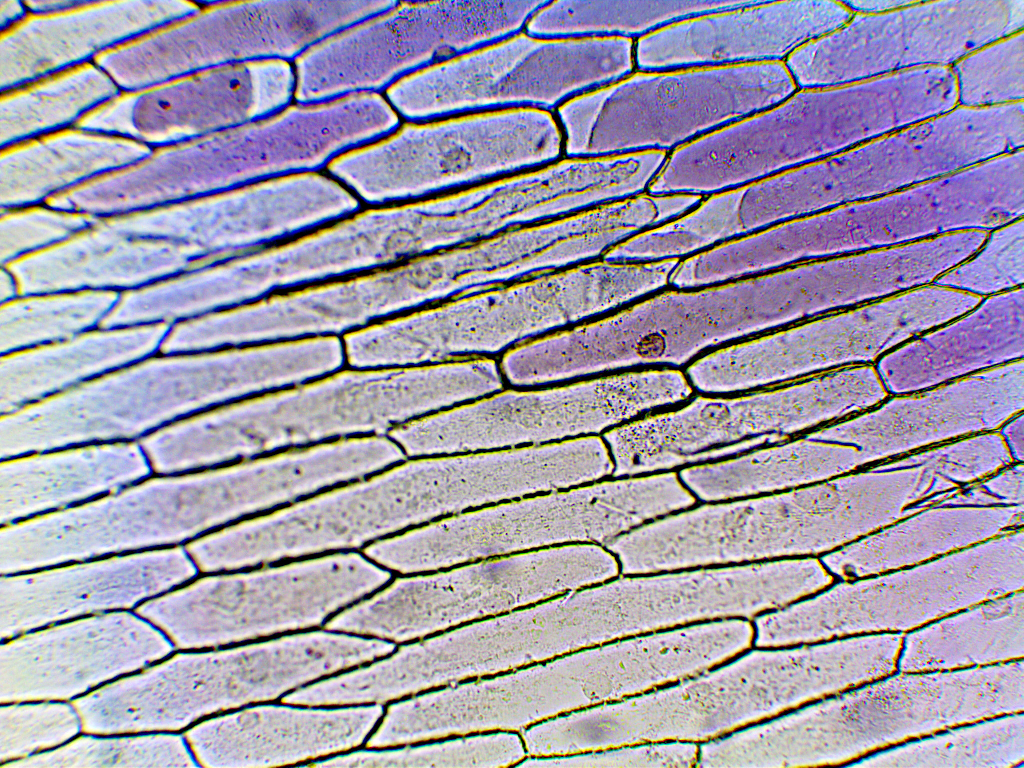
In this post, I will show how to make a wet mount slide for looking onion cells under a microscope.
Making the slide
- Take a clean slide and place a drop of water in the centre
- Take a small piece of onion and carefully peel the translucent membrane from the rough underside
of the slide. To peel the membrane, you can either use a sharp blade or a pair of tweezers. It is important to do this step carefully so as to not break too many cells. So, ideally always hold the peeled membrane at the edges. - Now carefully, place the membrane in the drop of water placed earlier on the slide.
- You may want to put a small drop of tincture iodine over the onion membrane. This is to help create contrast between cell nuclei and other parts of cells.
- Finally, gently lower a cover slip over the membrane.
Micrographs
Below are the micrographs of the onion cells. The nuclei are the small dark circles and the thick black lines are the cell walls.


helpfull , buh i wanted more